Homework 3 and Mid-term
ECE 4339 Spring
2014
Han Q. Le
(copyrighted) U. of Houston
Choose a combination of problems so that the total
>= 100 pts to submit as part 1.
Then the rest can be submitted a week later as part 2. Must follow
numerical order in each part.
1. (15 pts) Basic review: frequency, photon quantum energy, and units.
1.1 (2 pts) Question 1: speed of light
The speed of light c is ![]() meter/sec. Calculate
the speed of light in the following units: cm/sec; μm/ps; where:
meter/sec. Calculate
the speed of light in the following units: cm/sec; μm/ps; where: ![]() . Try to remember the
value of c
in μm/ps. It is a very useful unit.
. Try to remember the
value of c
in μm/ps. It is a very useful unit.
1.2 (2 pts) Question 2: frequency
Calculate the frequency of light in unit of THz
(Terahertz), which is ![]() Hz, using the result
of c
in the unit of μm/ps from question 1 above, for the following
wavelengths: 0.78 μm (the wavelength of CD laser), 0.65 μm (the
wavelength of DVD laser), 0.633 μm (HeNe laser wavelength), 0.532
μm (the wavelength of doubled Nd:YAG, such as green laser pointer
), and 0.405 μn (BluRay laser). What is the color of each
wavelength? (search for picts that shows colors of these
wavelength on the Internet).
Hz, using the result
of c
in the unit of μm/ps from question 1 above, for the following
wavelengths: 0.78 μm (the wavelength of CD laser), 0.65 μm (the
wavelength of DVD laser), 0.633 μm (HeNe laser wavelength), 0.532
μm (the wavelength of doubled Nd:YAG, such as green laser pointer
), and 0.405 μn (BluRay laser). What is the color of each
wavelength? (search for picts that shows colors of these
wavelength on the Internet).
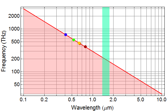
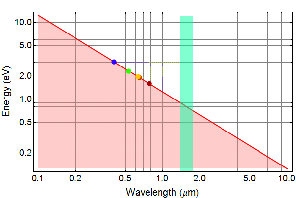
1.3 (1 pt) Question 3: λ vs. f
Plot lightwave frequency (in unit of THz) as a function of wavelength (in unit of μm) on log-log scale for λ (wavelength) from 10 μm to 0.1 μm.
1.3 Answer given (but you must import and paste in your work - unless you use Mathematica)
1.4 (2 pts) Question 4: quantum
We discuss Planck’s light quantum, the smallest
anount of energy that a light carries, as: E=h ν
where h is Planck’s constant and ν is light frequency. (h= 6.62606896 ![]() Joule/s). Find the
photon energy for each light in question 2.
Joule/s). Find the
photon energy for each light in question 2.
1.5 (2 pts) Question 5: Unit of eV
A most useful unit of energy is eV= electron-Volt:
it is the energy to move an electron across a potential of 1 V.
What is one eV in the unit of Joule. (note the charge of an
electron is: e=1.602176487 ![]() Coulomb.
Coulomb.
1.6 (2 pts) Question 6: photon energy
What is the energy of each photon in question 5 in unit of eV?
1.7 (1 pt) Question 7: photon energy vs. λ
Plot the photon energy (in unit eV) as a function of wavelength on log-log scale for λ (wavelength) from 10 μm to 0.1 μm.
1.7 Answer (but you must import and paste in your work - unless you use Mathematica)
1.8 (3 pts) Question 8: astronomical photon energy
This is an exercise in unit conversion. Below is the plot of gamma ray intensity as function of photon energy from the center of our galaxy (Milkyway) obtained by Max Planck Institute for Astrophysics. (γ-ray is photon with very high energy.)
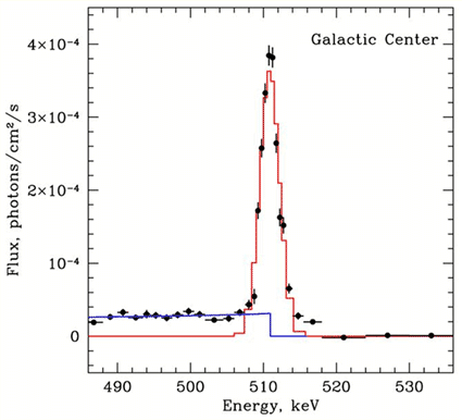
Using the formula: ![]() , and the electron rest
mass is 9.10938215
, and the electron rest
mass is 9.10938215![]() Kg, guess what nature
of the peak at 511 keV (kilo eV) in the graph is. Calculate the
511-keV-line intensity (the peak if the graph) in unit of
Kg, guess what nature
of the peak at 511 keV (kilo eV) in the graph is. Calculate the
511-keV-line intensity (the peak if the graph) in unit of ![]() Hint, look up
matter-antimatter, and electron its anti-matter.
Hint, look up
matter-antimatter, and electron its anti-matter.
2. (25 pts) Carrier density and Fermi level in GaAs
2.1 (5 pts)
Go to lecture: http://www0.egr.uh.edu/courses/ece/ece4339-4119/Class%20 Notes/Chapter%203 %20-%20 Carriers %20 in %20 semiconductor %20-Part %202.html . See calculation of 6.3.1 (the default parameters are for GaAs).
For T=300K, vary the conduction band Fermi level
from -0.2 eV to 0.2 eV (the value is relative to the conduction
band edge) in 5 steps (your choice of the steps, but the 2
endpoints have to be -0.2 and 0.2 eV, for example: -0.2, -0.15 , 0
,0.75, 0.2 etc... don’t follow this example). Record the
calculated the carrier density. Then, use the approximated
formula:
![]() which
was already given for GaAs: (cf. Chapter 3, 7.1)
which
was already given for GaAs: (cf. Chapter 3, 7.1)
![]()
to calculate the carrier densities for the same Fermi levels. Plot
the carrier density by both approaches vs Fermi levels on
Log-Linear scale. (Carrier density on log scale, Fermi level on
linear scale). What do you think
of the approximated formula? (Is it reasonable?)
2.2 (4 pts)
Do the same as above for T=500 K. But here, you don’t have to do the part with approximated formula. Make a log-linear plot of carrier density for both 300 K you find in 1.1 and 500 K you find here.
2.3 (4 pts)
Let a GaAs crystal be Te-doped at ![]()
![]() (and no other
dopants). At 300K, all Te donors are ionized, in other words, all
Te-electrons are free. Find the Fermi level relative to the
conduction band. (You can use the web lecture calculator and if
so, no need to show your calculation, but show a screen view that
you did it - it is also OK to do your own calculation).
(and no other
dopants). At 300K, all Te donors are ionized, in other words, all
Te-electrons are free. Find the Fermi level relative to the
conduction band. (You can use the web lecture calculator and if
so, no need to show your calculation, but show a screen view that
you did it - it is also OK to do your own calculation).
2.4 (4 pts)
Do the same as in 1.1 but for holes and use section
6.3.3 calculation. Vary the Fermi level from 0.2 to -0.1 eV
(relative to the valence band edge). (You must calculate ![]() ). Plot the carrier
density vs -Fermi (not Fermi, just change the sign to get -Fermi
to plot, in other words, if Fermi valence is -0.1 eV, phot the
point as +0.1 eV).
). Plot the carrier
density vs -Fermi (not Fermi, just change the sign to get -Fermi
to plot, in other words, if Fermi valence is -0.1 eV, phot the
point as +0.1 eV).
2.5 (4 pts)
Plot the electron density you find in 1.1 (neglect the approximation formula results) and the hole density found in 1.4 vs Fermi Level on the same graph and compare (note: the Fermi level will be kept the same sign for electrons, and reversed sign for holes). Discuss why carrier density for holes is so much larger than electrons for the same Fermi level.
2.6 (4 pts)
Another GaAs crystal layer is Mg-doped at ![]()
![]() (and no other
dopants). This layer and the layer in 1.3 are joined together to
form a junction and at thermal equilibrium. Draw the conduction
and valence bands of each layer relative to each other. (For this
question, you need not be concerned with what happens at the
interface region between the 2 layers. Just plot the band
structure of one layer on the right and that of the other layer on
the left (see lecture Chapter 3, section 9.1)
(and no other
dopants). This layer and the layer in 1.3 are joined together to
form a junction and at thermal equilibrium. Draw the conduction
and valence bands of each layer relative to each other. (For this
question, you need not be concerned with what happens at the
interface region between the 2 layers. Just plot the band
structure of one layer on the right and that of the other layer on
the left (see lecture Chapter 3, section 9.1)
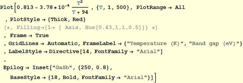
3. (15 pts) Intrinsic vs. doped carriers in GaSb
Refer to this web site: http://www.ioffe.ru/SVA/NSM/Semicond/
for various parameters of GaSb. Use the calculation in Chapter 3, 6.3.4,
but make sure with GaSb parameters for the following questions.
Neglect the light hole, use only the heavy hole effective mass.
For band gap dependence of temperature, use the formula:
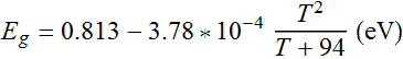
This is a plot for your reference:
3.1 Question 1 (5 pts)
Find the intrisic carrier density and Fermi level at T=100, 200, 300, 400, 500 K. Then plot the intrinsic Fermi level as a function of temperature. Use the midgap as the reference point. (see 6.4.1 as an example).
3.2 Question 2 (4 pts)
Plot the intrisic carrier density obtained in 2.1 vs. temperature on a log-linear scale. Discuss.
3.3 Question 3 (4 pts)
Let T=300 K. Plot the carrier density product ![]() as a function of Fermi
level from -0.4 to 0.4 eV on a log-linear scale with a minimum of
6 values of Fermi level. What do you observe? Discuss.
as a function of Fermi
level from -0.4 to 0.4 eV on a log-linear scale with a minimum of
6 values of Fermi level. What do you observe? Discuss.
3.4 Question 4 (2 pts)
An GaSb crystal is Mg-doped with ![]() . Find the minority carrier concentration
at 300 K.
. Find the minority carrier concentration
at 300 K.
4. (20 pts) Carriers relaxation and luminescence
4.1 (2 pts)
When electrons and holes are injected or generated in a semiconductor with energy much higher above conduction band and valence band, respectively, what will happen to them? and draw a diagram to explain. Note: by convention for holes, the energy scale is inverted, in other words, if a hole with energy that appears to be below the valence band edge in a diagram, it means its energy is high above the valence band edge. (Think of it as the mirror image of electron energy diagram across the horizontal line).
4.2 (2 pts)
When electrons and holes relax to the bottom of conduction bands, what relaxation process shown in the class demonstration?
4.3 (4 pts)
In the class demonstration, when the red (650 nm) laser light is shone of various materials, only scattered laser light of the same color was seen. But when the blue laser (405 nm) was used, lights of different colors appeared: greenish, yellowish, orange, even white (with a tint of blue that was from the original laser light). Did the laser light change its color? Explain the phenomenon. Why did it happen only with the blue laser but not the red laser (Remember what discussed in class). Explain if the following statement is true or false and why: The observed colors, (greenish, yellowish, orange, white) is a function of both the blue laser light and the material properties; if the blue laser wavelength is changed to, for example, 390 nm instead of 405 nm, the emitting color will also change.
4.4 (4 pts)
![]() is an alloy of GaAs
and AlAs, of which the direct bandgap is given by the formula:
is an alloy of GaAs
and AlAs, of which the direct bandgap is given by the formula:
![]()
Let ![]() (at 300 K), Plot the
bandgap
(at 300 K), Plot the
bandgap ![]() as a function of
alloy fraction x, for x from 0 to 0.4
as a function of
alloy fraction x, for x from 0 to 0.4
4.5 (3 pts)
A person has 2 wafers with an epitaxial layer of ![]() on it with two
different values of x.
But he/she doesn’t remember what the x values are, and cannot tell which and which.
If you have a blue laser and an optical spectrum analyzer (which
is also called a spectrometer, an instrument that can measure the
spectrum of light - you can look it up and read more), what will
be a simple measurement you can do to help the person determine x and tell which and which
wafer? (Hint: see the next question). Do not just use a name for
the experiment, you must also identify the physical process and
explain why your experiment can help solving the problem.
on it with two
different values of x.
But he/she doesn’t remember what the x values are, and cannot tell which and which.
If you have a blue laser and an optical spectrum analyzer (which
is also called a spectrometer, an instrument that can measure the
spectrum of light - you can look it up and read more), what will
be a simple measurement you can do to help the person determine x and tell which and which
wafer? (Hint: see the next question). Do not just use a name for
the experiment, you must also identify the physical process and
explain why your experiment can help solving the problem.
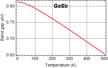
4.6 (5 pts)
The experiment was done at liquid ![]() temperature, T=77 K,
and you obtained 2 spectra below: wafer A gave the left spectrum
and wafer B gave the right spectrum. Determine the Al x concentration for each
wafer, and describe the emission color from each wafer (e. g. red,
green,...). You are welcome to find online the closest hue for
each wavelength and submit for extra credit. You can also obtain a
graph of the visible spectrum vs wavelength and use arrows to
point to the color of the luminescence from each wafer.
temperature, T=77 K,
and you obtained 2 spectra below: wafer A gave the left spectrum
and wafer B gave the right spectrum. Determine the Al x concentration for each
wafer, and describe the emission color from each wafer (e. g. red,
green,...). You are welcome to find online the closest hue for
each wavelength and submit for extra credit. You can also obtain a
graph of the visible spectrum vs wavelength and use arrows to
point to the color of the luminescence from each wafer.

To solve this problem, you should use this
calculation of luminescence spectrum in lecture:
http://www0.egr.uh.edu/courses/ece/ece4339-4119/Class%20Notes/Chapter%204%20-%20Excess%20carriers_html_Part1.html
in section 2.2.2. However, note that the plot there is
luminescence vs. photon energy (eV), not wavelength, hence you
must convert wavelength to photon energy. Make sure you set the
temperature at 77 K. Vary x concentration such that you obtain a
spectrum with the peak emission photon energy best matched the
measured spectra above. You should obtain x with an accuracy better
than 5% for full credit.
5. (25 pts) Mobility and diffusion illustration
As we learn, excess carriers diffuse and at the
same time decay via recombination, resulting in an effective
“spread distance” called diffusion length: ![]() where τ is the
carrier lifetime, D is
the diffusion coefficient or diffusivity for short, which is
related to the mobility by the Einstein relation:
where τ is the
carrier lifetime, D is
the diffusion coefficient or diffusivity for short, which is
related to the mobility by the Einstein relation:
![]()
The larger L
is, the farther do carriers spread out.
In the following, refer to Chapter 4, part 2, Section 5.4
5.1 (5 pts) Consider example 5.4.1 of Chapter 4
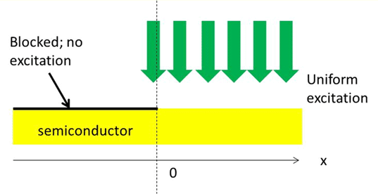
Set P=2, μ=1, Find the values of ![]() and
and ![]() such that you have a
physically valid solution. Copy and paste the 3 plots: carrier
density, carrier flux, and light emission intensity. If you obtain
a proof for what
such that you have a
physically valid solution. Copy and paste the 3 plots: carrier
density, carrier flux, and light emission intensity. If you obtain
a proof for what ![]() and
and ![]() should be as a
function of pump rate P, you will get extra credit.
should be as a
function of pump rate P, you will get extra credit.
5.2 (5 pts) Vary μ
Obtain the image of light emission intensity for μ=0.1 and 5. Compare the two cases and discuss how they are different
5.3 (5 pts) Consider example 5.4.2 of Chapter 4
Do the same thing as 5.1, and set a=1. Obtain the
value of ![]() ,
, ![]() ,
, ![]() and
and ![]() (hint,
(hint, ![]() =
= ![]() and
and ![]() ).
).
5.4 (5 pts) Larger gap
Do the same as above with a=4. Compare the peak carrier density for the case in 5.3 and 5.4 and discuss their difference (why the peak carrier population density for a=4 is larger than that for a=1?)
5.5 (5 pts) Vary μ
Let a=1, obtain results for 2 cases: μ=0.001 and 5. (for μ=0.001, just type in the number, the slider won’t go lower than 0.1). Note what the peak carrier density is for μ=0.001 (it is essentially the same as the pump rate), then compare the peak carrier density again for μ=5. Explain why so in simple terms. (Use your intuition, if you have the same amount of money but spread out more, how much is left?).
6. (50 pts) Techical reading and analytical interpretation
Read the paper: “Time-resolved imaging of radiative recombination in 4H-SiC p-i-n diode” by A. Galeckas et al., Appl. Phys. Lett. Vol. 74, pp. 3398-3400, 1999. It is available on the last section of lecture Chapter 4, part 2.
To answer the questions below, you need to read the paper. Looking at a figure alone will NOT be enough. Read those paragraphs pertaining to each figure to answer them. You do NOT need to understand 100% of what in the paper to answer all these questions below.
5 points each question.
6.1
Label on Fig. 1 (copy and use some software such as ppt drawing to show) where the substrate is, the epilayers and their thickness, the mesa and its dimension.
6.2
What does figure 2 describe? What color do you think it looks like?
6.3
look at Fig. 3. Look at the black-white inset photo. What is the bright streak at the top? Why does it get darker as you go to the bottom?
6.4
Look at Fig. 3 again. What does it plot? Is the
vertical axis on log scale or linear scale? If we assume that the
emitting light intensity is proportional to the electron-hole (EH)
density, how do you express the spatial variation of the EH
density along these layers? Look at the curve for 100 A/cm2, if
you approximate the carrier density profile as ![]() where x is the
distance and
where x is the
distance and ![]() is the diffusion
length, what are the diffusion length values for n- and n+ region?
is the diffusion
length, what are the diffusion length values for n- and n+ region?
6.5
Recalling that diffusion length ![]() where D is the
diffusivity and τ is the effective carrier lifetime, let’s assume
that the D is the diffusivity is approximately the same for both
n- and n+ region, which region, n- or n+, has a longer carrier
lifetime? Can you speculate why so?
where D is the
diffusivity and τ is the effective carrier lifetime, let’s assume
that the D is the diffusivity is approximately the same for both
n- and n+ region, which region, n- or n+, has a longer carrier
lifetime? Can you speculate why so?
6.6
Recalling that the diffusivity D is also proportional to the mobility. Which region, n- or n+ do you think the mobility is higher? Do you think that both mobility and carrier lifetime contribute to the difference in the diffusion length between the two regions?
6.7
Look at Fig. 4. What does the top photo (Fig. 4(a)) show? Copy and paste the figure and draw a arrow to show where you think there is an excess electron-hole population. Do you think that this is a spatially uniform diode along the horizontal direction? In Fig. 4(a), which part, left or right, is more efficient as an LED? What does Fig. 4(b) show? Is the vertical scale logarithmic or linear?
6.8
Look at Fig. 5. What do the researchers claim the effective carrier lifetime they measured for each region, n- and n+? How did they measure these values?
6.9
Go back to questions (6.4) and (6.5), use the lifetime given in the paper you that you find in question (6.8) above, use the value of diffusion length you find in (6.4), infer what you think the diffusivity for each region are, using appropriate relations.
6.10
Comment on what you learn from this paper
7. (50 pts) Real life application
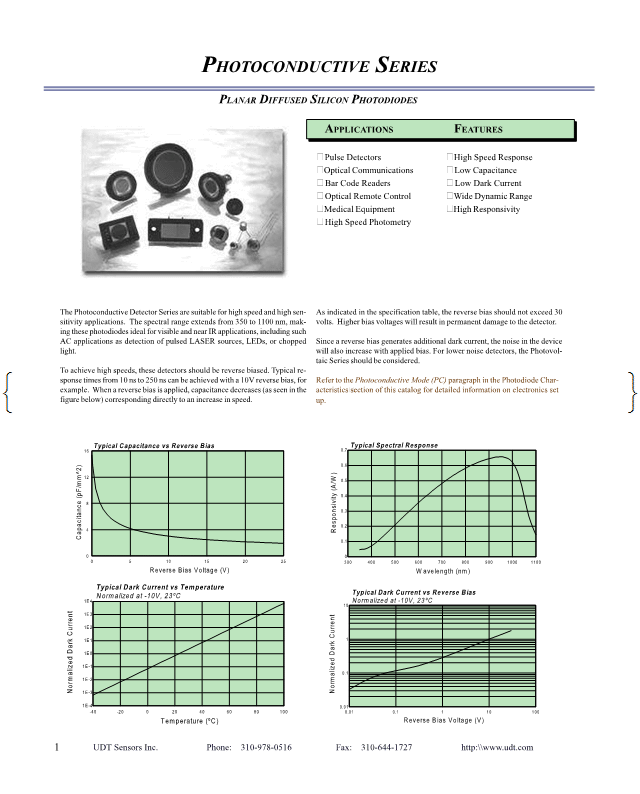
Suppose you want to build something that requires an optical detector. Attached at the end is a technical brochure from a company selling Si detectors. The purpose of this problem is to ask you to read, apply what you learn to understand, so that you will be an informed user of these products.
7.1 (5 pts)
In a few words, summarize: What does the brochure describe? Explain in simple words how these detectors work. If you have 1.5-μm laser (it means the laser wavelength is 1.5 μm), would you buy these detectors to detect your laser light? (hint: read the brochure carefully)
7.2 (10 pts)
The brochure mentions these features:
- low capacitance: explain what good is low
capacitance?
- low dark current: what is dark current
and what good is dark current? (remember discussion in class)
- high responsivity: what is responsivity?
What is its unit? Why high responsivity is good?
Extra note: The function of a photodetector (PD) is to detect light (photons). When a photon whose energy is above a semiconductor bandgap impinges on the semiconductor and is absorbed, it generates an electron hole pair, which can be collected with electrodes. The merit of a PD is a measure that determines how strong it can give a current signal for a given light power input. Think of this: What is the light input unit? What is the detector output unit? How would one determine the proportionality between the output vs. input? This proportionality coefficient is called responsivity. If a detector gives 1 mA output current for 3-mW light input, what is its responsivity? If a detector gives 0.1 mA current output without any light input- i. e. in total darkness, is this 0.1 mA signal useful? can it be detrimental?)
7.3 (10 pts)
Look at the chart labeled “Typical Dark Current vs Temperature”. Write an expression that describes the relationship of dark current (relative unit only) and temperature. Suppose you plan to use this detector in a system that detects the ambient light level around a car and automatically turns on the car headlight if it senses darkness. For winter in Bismarck, North Dakota (-40 C) this system typically has a signal current/dark current ratio of 1000 during bright sunshine daytime. You bring this system to Houston for the summer (~ 80 C under the hood of a car with engine on). What is your signal current/dark current ratio now? Do you think the circuit will work reliably?
7.4 (15 pts)
Review Chapter 3, Section 7.5.1 about intrinsic
carrier density in Si as a function of temperature. This detector
has an intrinsic drift region sandwiched between lightly doped
regions. Use the same method in 7.5.1, calculate the intrinsic
carrier concentration for temperature from -40 C to 100 C. Then,
calculate and plot the drift current density as a function of
temperature:
![]()
where symbol n and p represent electron and hole
density respectively.
Plot the drift current in relative unit (no need for absolute
unit). If you wish, you can assume for simplicity that mobility is
constant vs. temperature and carrier density. (Of course, we learn
in HW 1 that mobility does depends on temperature, but just
neglect it here for approximation). Compare your plot
qualitatively with the data on dark current vs. temperature (Note:
the calculation for realistic dark current is a bit more
complicated with impurity effects and leakage current included).
7.5 (10 pts)
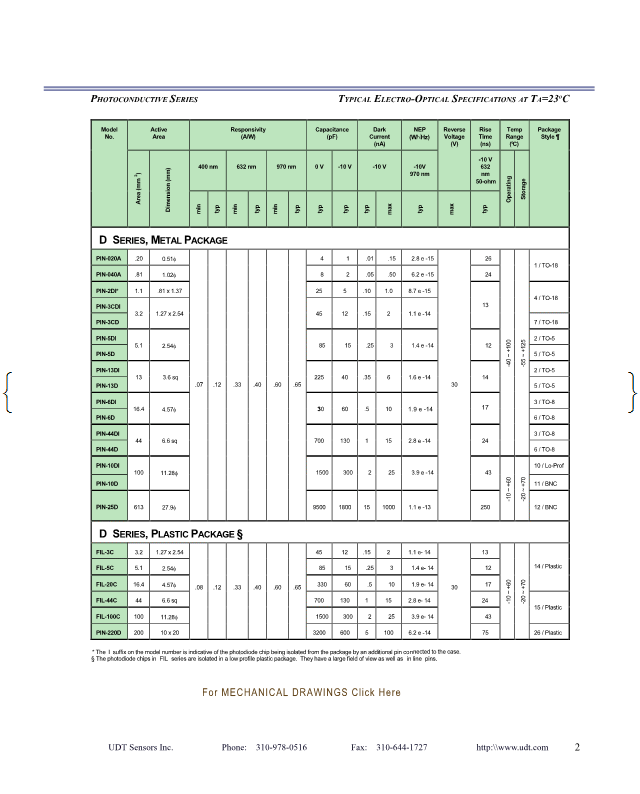
Look at the data for D-series detectors, metal
package. Plot (scatter plot) the detector typical dark current vs.
the detector active area (Active area of a detector is the area
that responds to light). Any comments on the relationship? Why
would you want large area detector if the dark current is higher?
Technical brochure